Ankylosing spondylitis

Ankylosing spondylitis (AS) is a type of arthritis in which there is a long-term inflammation of the joints of the spine. Typically the joints where the spine joins the pelvis are also affected. Occasionally other joints such as the shoulders or hips are involved. Eye and bowel problems may also occur. Back pain is a characteristic symptom of AS, and it often comes and goes. Stiffness of the affected joints generally worsens over time.
Although the cause of ankylosing spondylitis is unknown, it is believed to involve a combination of genetic and environmental factors. More than 85% of those affected in the UK have a specific human leukocyte antigen known as the HLA-B27 antigen. The underlying mechanism is believed to be autoimmune or autoinflammatory. Diagnosis is typically based on the symptoms with support from medical imaging and blood tests. AS is a type of seronegative spondyloarthropathy, meaning that tests show no presence of rheumatoid factor (RF) antibodies. It is also within a broader category known as axial spondyloarthritis.
There is no cure for ankylosing spondylitis. Treatments may improve symptoms and prevent worsening. This may include medication, exercise, physical therapy, surgery in rare cases. Medications used include NSAIDs, steroids, DMARDs such as sulfasalazine, and biologic agents such as TNF inhibitors.
Between 0.1% and 0.8% of people are affected. Onset is typically in young adults. Males and females are equally affected. It used to be thought that three times as many men as women had the disease. This was based on a diagnosis of the disease using x-ray. Men are more likely than women to experience changes to the bones and fusion, and thus they were being picked up using x-ray.
Over time MRIs were developed which could identify inflammation. Women are more likely than men to experience inflammation rather than fusion. The condition was first fully described in the late 1600s by Bernard Connor, but skeletons with ankylosing spondylitis are found in Egyptian mummies. The word is from Greek ankylos meaning crooked, curved or rounded, spondylos meaning vertebra, and -itis meaning inflammation.
As the disease progresses, loss of spinal mobility and chest expansion, with a limitation of anterior flexion, lateral flexion, and extension of the lumbar spine, are seen. Systemic features are common, with weight loss, fever, or fatigue often present. Pain is often severe at rest but may improve with physical activity, but inflammation and pain to varying degrees may recur regardless of rest and movement.
AS can occur in any part of the spine or the entire spine, often with pain referred to one or the other buttock or the back of the thigh from the sacroiliac joint. Arthritis in the hips and shoulders may also occur. When the condition presents before the age of 18, it is more likely to cause pain and swelling of large lower limb joints, such as the knees. In prepubescent cases, pain and swelling may also manifest in the ankles and feet where heel pain and enthesopathy commonly develop. Less commonly ectasia of the sacral nerve root sheaths may occur.
About 30% of people with AS will also experience anterior uveitis, causing eye pain, redness, and blurred vision. This is thought to be due to the association that both AS and uveitis have with the inheritance of the HLA-B27 antigen. Cardiovascular involvement may include inflammation of the aorta, aortic valve insufficiency or disturbances of the heart's electrical conduction system. Lung involvement is characterized by progressive fibrosis of the upper portion of the lung.
Single nucleotide polymorphism (SNP) A/G variant rs10440635 close to the PTGER4 gene on human chromosome 5 has been associated with an increased number of cases of ankylosing spondylitis in a population recruited from the United Kingdom, Australia, and Canada. The PTGER4 gene codes for the prostaglandin EP4 receptor (EP4), one of four receptors for prostaglandin E2. Activation of EP4 promotes bone remodeling and deposition (see EP4, bone) and EP4 is highly expressed at vertebral column sites involved in ankylosing spondylitis. These findings suggest that excessive EP4 activation contributes to pathological bone remodeling and deposition in ankylosing spondylitis and that the A/G variant rs10440635a of PTGER4 predisposes to this disease, possibly by influencing EP4's production or expression pattern.
The association of AS with HLA-B27 suggests the condition involves CD8 T cells, which interact with HLA-B. This interaction is not proven to involve a self-antigen, and at least in the related reactive arthritis, which follows infections, the antigens involved are likely to be derived from intracellular microorganisms. There is, however, a possibility that CD4+ T lymphocytes are involved in an aberrant way, since HLA-B27 appears to have a number of unusual properties, including possibly an ability to interact with T cell receptors in association with CD4 (usually CD8+ cytotoxic T cell with HLAB antigen as it is a MHC class 1 antigen).
"Bamboo spine" develops when the outer fibers of the fibrous ring (anulus fibrosus disci intervertebralis) of the intervertebral discs ossify, which results in the formation of marginal syndesmophytes between adjoining vertebrae.
While ankylosing spondylitis can be diagnosed through the description of radiological changes in the sacroiliac joints and spine, there are currently no direct tests (blood or imaging) to unambiguously diagnose early forms of ankylosing spondylitis (non-radiographic axial spondyloarthritis). Diagnosis of non-radiologic axial spondyloarthritis is therefore more difficult and is based on the presence of several typical disease features.
These diagnostic criteria include:
Inflammatory back pain:
• Chronic, inflammatory back pain is defined when at least four out of five of the following parameters are present: (1) Age of onset below 40 years old, (2) insidious onset, (3) improvement with exercise, (4) no improvement with rest, and (5) pain at night (with improvement upon getting up)
• Past history of inflammation in the joints, heels, or tendon-bone attachments
• Family history for axial spondyloarthritis or other associated rheumatic/autoimmune conditions
• Positive for the biomarker HLA-B27
• Good response to treatment with nonsteroidal anti-inflammatory drugs (NSAIDs)
• Signs of elevated inflammation (C-reactive protein and erythrocyte sedimentation rate)
• Manifestation of psoriasis, inflammatory bowel disease, or inflammation of the eye (uveitis)
• If these criteria still do not give a compelling diagnosis magnetic resonance imaging (MRI) may be useful. MRI can show inflammation of the sacroiliac joint.
The Bath Ankylosing Spondylitis Functional Index (BASFI) is a functional index which can accurately assess functional impairment due to the disease, as well as improvements following therapy. The BASFI is not usually used as a diagnostic tool, but rather as a tool to establish a current baseline and subsequent response to therapy.
The mainstay of therapy in all seronegative spondyloarthropathies are anti-inflammatory drugs, which include NSAIDs such as ibuprofen, phenylbutazone, diclofenac, indomethacin, naproxen and COX-2 inhibitors, which reduce inflammation and pain. 2012 research showed that those with AS and elevated levels of acute phase reactants seem to benefit most from continuous treatment with NSAIDs.
Medications used to treat the progression of the disease include the following:
Disease-modifying antirheumatic drugs (DMARDs) such as sulfasalazine can be used in people with peripheral arthritis. For axial involvement, evidence does not support sulfasalazine. Other DMARDS, such as methotrexate, did not have enough evidence to prove their effect. Generally, systemic corticosteroids were not used due to lack of evidence. Local injection with corticosteroid can be used for certain people with peripheral arthritis.
Tumor necrosis factor-alpha (TNFα) blockers (antagonists), such as the biologics etanercept, infliximab, golimumab and adalimumab, have shown good short-term effectiveness in the form of profound and sustained reduction in all clinical and laboratory measures of disease activity. Trials are ongoing to determine their long-term effectiveness and safety. The major drawback is the cost. An alternative may be the newer, orally-administered non-biologic apremilast, which inhibits TNF-α secretion, but a recent study did not find the drug useful for ankylosing spondylitis.
Anti-interleukin-6 inhibitors such as tocilizumab, currently approved for the treatment of rheumatoid arthritis, and rituximab, a monoclonal antibody against CD20, are also undergoing trials.
Interleukin-17A inhibitor secukinumab is an option for the treatment of active ankylosing spondylitis that has responded inadequately to (TNFα) blockers.
• Low intensity aerobic exercise
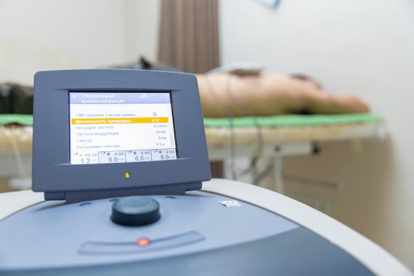
• Transcutaneous electrical nerve stimulation (TENS)
• Thermotherapy
• Proprioceptive neuromuscular facilitation (PNF)
• Exercise programs, either at home or supervised
• Hydrotherapy
• Group exercises; e.g., Pilates
Moderate-to-high impact exercises like jogging are generally not recommended or recommended with restrictions due to the jarring of affected vertebrae that can worsen pain and stiffness in some with AS.
Osteoporosis is common in ankylosing spondylitis, both from chronic systemic inflammation and decreased mobility resulting from AS. Over a long-term period, osteopenia or osteoporosis of the AP spine may occur, causing eventual compression fractures and a back "hump". Hyperkyphosis from ankylosing spondylitis can also lead to impairment in mobility and balance, as well as impaired peripheral vision, which increases the risk of falls which can cause fracture of already-fragile vertebrae. Typical signs of progressed AS are the visible formation of syndesmophytes on X-rays and abnormal bone outgrowths similar to osteophytes affecting the spine. In compression fractures of the vertebrae, paresthesia is a complication due to the inflammation of the tissue surrounding nerves.
Organs commonly affected by AS, other than the axial spine and other joints, are the heart, lungs, eyes, colon, and kidneys. Other complications are aortic regurgitation, Achilles tendinitis, AV node block, and amyloidosis. Owing to lung fibrosis, chest X-rays may show apical fibrosis, while pulmonary function testing may reveal a restrictive lung defect. Very rare complications involve neurologic conditions such as the cauda equina syndrome.
As increased mortality in ankylosing spondylitis is related to disease severity, factors negatively affecting outcomes include:
• Male sex
• Plus 3 of the following in the first 2 years of disease:
• Erythrocyte sedimentation rate (ESR) >30 mm/h
• Unresponsive to NSAIDs
• Limitation of lumbar spine range of motion
• Sausage-like fingers or toes
• Oligoarthritis
• Onset <16 years old
The anatomist and surgeon Realdo Colombo described what could have been the disease in 1559, and the first account of pathologic changes to the skeleton possibly associated with AS was published in 1691 by Bernard Connor. In 1818, Benjamin Brodie became the first physician to document a person believed to have active AS who also had accompanying iritis.
In 1858, David Tucker published a small booklet which clearly described the case of Leonard Trask, who suffered from severe spinal deformity subsequent to AS. In 1833, Trask fell from a horse, exacerbating the condition and resulting in severe deformity. Tucker reported:
It was not until he [Trask] had exercised for some time that he could perform any labor ... [H]is neck and back have continued to curve drawing his head downward on his breast.
This account became the first documented case of AS in the United States, owing to its indisputable description of inflammatory disease characteristics of AS and the hallmark of deforming injury in AS.
In the late nineteenth century, the neurophysiologist Vladimir Bekhterev of Russia in 1893, Adolph Strümpell of Germany in 1897, and Pierre Marie of France in 1898 were the first to give adequate descriptions which permitted an accurate diagnosis of AS prior to severe spinal deformity. For this reason, AS is also known as Bekhterev disease, Bechterew's disease or Marie–Strümpell disease.
Source www.wikipedia.org
